


What is Atrial Fibrillation?
A common heart arrhythmia

Recently, a family member called me up in a panic. She had visited her regular doctor for a regular appointment and had an EKG performed. It showed atrial fibrillation. She knew that I liked to draw pictures to explain medical things simply, so asked me to help her understand what this meant. I asked her to flip to Page 32 of my book, Diagnosketch, and told her this:
The left picture shows a normal heart. Electrical impulses start in one spot and then travel down a pathway. This causes the heart to pump in a regular, predictable way. We call this a ‘sinus rhythm.’
The right picture shows atrial fibrillation. Here, multiple spots fire all at once. This causes an irregular beating of the heart. The heart ‘fibrillates’ (fibrillate definition: vibrates in an uncoordinated way). You can see atrial fibrillation on an EKG or a cardiac monitor.
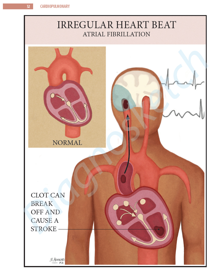
When you are in atrial fibrillation, your heart may not pump all of its blood out. Instead, some blood settles in the chambers of the heart. Clots can form. Sometimes, when a small clot is pumped out, it travels to the brain and blocks a blood vessel. This causes a stroke. Patients with atrial fibrillation often take blood thinners to prevent clots from forming so that they don’t develop a stroke.
After I explained this to my family member, she understood. I told her that many patients come to the ER with atrial fibrillation. They present with palpitations, chest pain, or shortness of breath. When we do an EKG, sometimes we find atrial fibrillation with a rapid heart rate. We give them medicines to slow their heart rates. Then, we talk about whether they need to start taking blood thinners.
And, on a side note, we often say ‘a-fib’ as a shortcut to atrial fibrillation.
Some facts about a-fib:
A-fib increases your risk of stroke by 5 times
15-20% of people who have strokes are found to have a-fib
2% of people younger than 65 years old have a-fib, but 9% of people older than 65 have it
*info from American Heart Association and CDC websites
Hope this was helpful.
Thanks for reading!